Home
Products
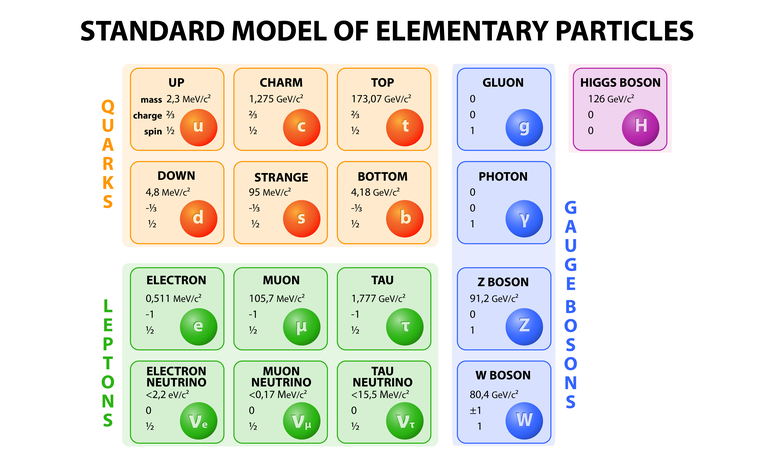
Physics research field


Type of experiment

Detector type

Behind the science
Tech in a nutshell
United Kingdom (EN)
Select your region or country.
Beam diagnostic & experiment
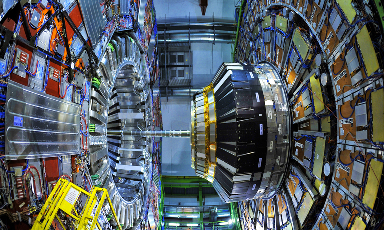
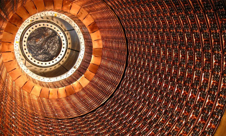
Particle accelerator facilities are widespread around the world, both for the study of particle physics and medical applications.
Beam diagnostic devices are key tools for the correct operation of accelerators, since they allow understanding of the beam properties and explain how waves behave in an accelerator. Beam diagnostics is a rich field; where a great variety of physical effects are made use of and imagination finds a wide playground. Today, there is a vast choice of diagnostic devices available, each employing different techniques. The beam parameters that should be monitored in an accelerator are usually: intensity, position, size and shape, emittance, energy and polarization. All of these measurements perturb the beam, some of these being negligible and others destructive.
It is common to say that an accelerator can never be better than its diagnostics and accurate equipment serves as a basis for this.
A particle accelerator can be operated with a high or low intensity beam. In the case of high intensity, it is possible to take direct measurements of the visible light emitted by the beam interaction with residual gas or fluorescent monitors.
Position measurements of the beam centroid in the beam pipe are of prime importance for the accelerator operation. On-line and non-interceptive measurements are required wherever possible. When the beam is in DC mode, a quantitative measurement of the beam centroid position can be done using a sCMOS camera.
In this case the beam excites the atoms of the residual gas in the beam pipe and they emit light. A sCMOS camera can sense this emitted light and its position can be deduced. Hamamatsu Photonics produces a range of sCMOS cameras designed particularly for this application.
The Cornell Electron-positron Storage Ring (CESR) is an electron-positron collider with a circumference of 768 meters, located 12 meters below the parking lot of the scenic Cornell University campus. It is capable of producing collisions between electrons and their anti-particles, positrons, with center-of-mass energies between 9 and 12 GeV. When an electron and positron collide and annihilate, the flash of energy results in the creation of new matter. This new matter, sometimes strange and exotic, is the main object of interest in the CLEO experiment.
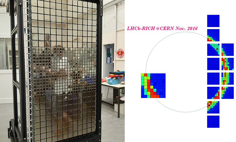
One of the main aims of beam diagnostics is to measure the longitudinal and vertical beam dynamics of single colliding bunches for luminosity optimisation. For the longitudinal beam diagnostics, the synchrotron light emitted both by electrons and positrons is reflected by a mirror into a streak camera. Hamamatsu Photonics C16910 Streak camera can measure electron bunch length with 800 fs time resolution.

sCMOS camera

Streak camera
Two PMT arrays are installed for electron/positron vertical beam size measurements. Each array has a 32 channel linear anode with an effective area per channel of 0.8x7mm. The PMT signal has a sub-nanosecond rise time that measures the individual vertical beam size of all the bunches in CESR.
Beam profile measurements exploit the residual-gas ionization profile measurement. The interaction between the beam and the background-residual gas creates electron-ion pairs that can be collected by a micro-channel plate (MCP) followed by a one-plane position sensitive device. As for beam centroid position measurements, the profile of the primary beam is deduced from the measurements of charges collected and amplified by the MCP.
Another technique for analyzing the beam profile measures the fluorescence of the residual gas interacting with the primary proton. The light in the visible range can be sensed by a sCMOS camera and allows for qualitative information on the beams size. In accelerators using this technique, special glass windows are used where the beam is intercepted by the monitor. The NIRS-HIMAC is a heavy ion synchrotron complex for medical use in Chiba, Japan. Precise control of position, intensity and shape of the beams is crucial in medical applications; where ion beams are used to irradiate tumours with millimetre precision or to produce large quantities of radioisotopes for diagnostics and therapy.
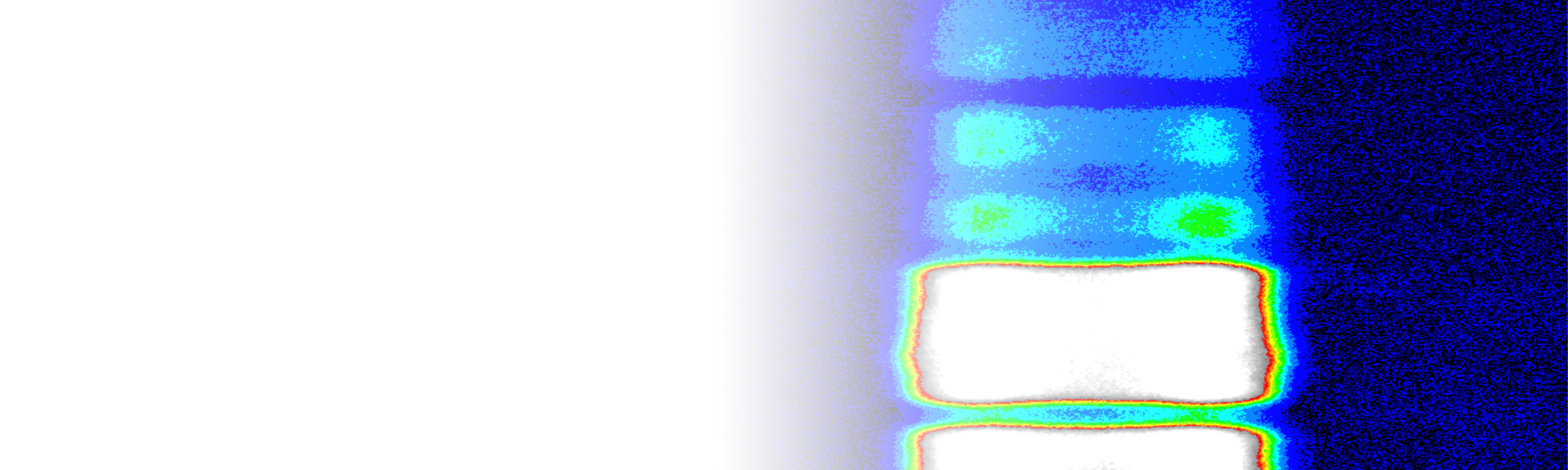
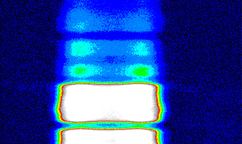
In this facility the beam profile monitoring system is composed by; a double stage MCP, an image intensifier and a sCMOS camera, as shown in the image below.

Ions generated by the beam at the inclined sheet beam target are collected with a radial electric field. This field is induced by semi-spherical electrodes and multiplied by a two stage MCP (Hamamatsu Photonics F2226: max. gain 1x10⁷). The luminescent light from the phosphor screen (decay time of the light : 100 ns at 1/10) is detected by a sCMOS camera, which is attached to an image intensifier (I.I.: max. gain 1x10⁴ : Hamamatsu Photonics C9546-04). An image of the beam profile at 8MeV and 5µA at the NIRS cyclotron is shown in the figure.
When the particle accelerator is operated with low intensity, in the case of radioactive ion beams for cancer therapy, the beam diagnostic device should have different characteristics. In this situation the diagnostic system is required to be very robust and cheap since it has to withstand beam tuning. This leads to long operating periods, human errors leading to overexposure, and all of the normal run operations.
This is why the adopted solution is usually a scintillation detector. It consists of a piece of scintillator material (usually NaI) optically coupled to a photosensor.
The scintillator intercepts the beam and produces an average of one photon per 100 eV of deposited energy. The light readout devices can be different depending on the purpose.
Photodiodes are better suited for current readout, photomultiplier tubes (PMTs) and avalanche photodiodes (APDs) are best for pulse counting and there are some PMTs which are calibrated specifically for single photon counting.
- Confirmation
-
It looks like you're in the . If this is not your location, please select the correct region or country below.
You're headed to Hamamatsu Photonics website for GB (English). If you want to view an other country's site, the optimized information will be provided by selecting options below.
In order to use this website comfortably, we use cookies. For cookie details please see our cookie policy.
- Cookie Policy
-
This website or its third-party tools use cookies, which are necessary to its functioning and required to achieve the purposes illustrated in this cookie policy. By closing the cookie warning banner, scrolling the page, clicking a link or continuing to browse otherwise, you agree to the use of cookies.
Hamamatsu uses cookies in order to enhance your experience on our website and ensure that our website functions.
You can visit this page at any time to learn more about cookies, get the most up to date information on how we use cookies and manage your cookie settings. We will not use cookies for any purpose other than the ones stated, but please note that we reserve the right to update our cookies.
1. What are cookies?
For modern websites to work according to visitor’s expectations, they need to collect certain basic information about visitors. To do this, a site will create small text files which are placed on visitor’s devices (computer or mobile) - these files are known as cookies when you access a website. Cookies are used in order to make websites function and work efficiently. Cookies are uniquely assigned to each visitor and can only be read by a web server in the domain that issued the cookie to the visitor. Cookies cannot be used to run programs or deliver viruses to a visitor’s device.
Cookies do various jobs which make the visitor’s experience of the internet much smoother and more interactive. For instance, cookies are used to remember the visitor’s preferences on sites they visit often, to remember language preference and to help navigate between pages more efficiently. Much, though not all, of the data collected is anonymous, though some of it is designed to detect browsing patterns and approximate geographical location to improve the visitor experience.
Certain type of cookies may require the data subject’s consent before storing them on the computer.
2. What are the different types of cookies?
This website uses two types of cookies:
- First party cookies. For our website, the first party cookies are controlled and maintained by Hamamatsu. No other parties have access to these cookies.
- Third party cookies. These cookies are implemented by organizations outside Hamamatsu. We do not have access to the data in these cookies, but we use these cookies to improve the overall website experience.
3. How do we use cookies?
This website uses cookies for following purposes:
- Certain cookies are necessary for our website to function. These are strictly necessary cookies and are required to enable website access, support navigation or provide relevant content. These cookies direct you to the correct region or country, and support security and ecommerce. Strictly necessary cookies also enforce your privacy preferences. Without these strictly necessary cookies, much of our website will not function.
- Analytics cookies are used to track website usage. This data enables us to improve our website usability, performance and website administration. In our analytics cookies, we do not store any personal identifying information.
- Functionality cookies. These are used to recognize you when you return to our website. This enables us to personalize our content for you, greet you by name and remember your preferences (for example, your choice of language or region).
- These cookies record your visit to our website, the pages you have visited and the links you have followed. We will use this information to make our website and the advertising displayed on it more relevant to your interests. We may also share this information with third parties for this purpose.
Cookies help us help you. Through the use of cookies, we learn what is important to our visitors and we develop and enhance website content and functionality to support your experience. Much of our website can be accessed if cookies are disabled, however certain website functions may not work. And, we believe your current and future visits will be enhanced if cookies are enabled.
4. Which cookies do we use?
There are two ways to manage cookie preferences.
- You can set your cookie preferences on your device or in your browser.
- You can set your cookie preferences at the website level.
If you don’t want to receive cookies, you can modify your browser so that it notifies you when cookies are sent to it or you can refuse cookies altogether. You can also delete cookies that have already been set.
If you wish to restrict or block web browser cookies which are set on your device then you can do this through your browser settings; the Help function within your browser should tell you how. Alternatively, you may wish to visit www.aboutcookies.org, which contains comprehensive information on how to do this on a wide variety of desktop browsers.
5. What are Internet tags and how do we use them with cookies?
Occasionally, we may use internet tags (also known as action tags, single-pixel GIFs, clear GIFs, invisible GIFs and 1-by-1 GIFs) at this site and may deploy these tags/cookies through a third-party advertising partner or a web analytical service partner which may be located and store the respective information (including your IP-address) in a foreign country. These tags/cookies are placed on both online advertisements that bring users to this site and on different pages of this site. We use this technology to measure the visitors' responses to our sites and the effectiveness of our advertising campaigns (including how many times a page is opened and which information is consulted) as well as to evaluate your use of this website. The third-party partner or the web analytical service partner may be able to collect data about visitors to our and other sites because of these internet tags/cookies, may compose reports regarding the website’s activity for us and may provide further services which are related to the use of the website and the internet. They may provide such information to other parties if there is a legal requirement that they do so, or if they hire the other parties to process information on their behalf.
If you would like more information about web tags and cookies associated with on-line advertising or to opt-out of third-party collection of this information, please visit the Network Advertising Initiative website http://www.networkadvertising.org.
6. Analytics and Advertisement Cookies
We use third-party cookies (such as Google Analytics) to track visitors on our website, to get reports about how visitors use the website and to inform, optimize and serve ads based on someone's past visits to our website.
You may opt-out of Google Analytics cookies by the websites provided by Google:
https://tools.google.com/dlpage/gaoptout?hl=en
As provided in this Privacy Policy (Article 5), you can learn more about opt-out cookies by the website provided by Network Advertising Initiative:
http://www.networkadvertising.org
We inform you that in such case you will not be able to wholly use all functions of our website.
Close